How can we help you?
Please get in contact with us to find out more about ECFP and whether we can help you.
ECFP has access to the cryo-FIB SEM facility located within the School of Physics & Astronomy at the University of Edinburgh. It provides a powerful three-in-one imaging facility for opaque soft matter. The instrument’s cryo mode gives the option of imaging a wide range of materials including liquids, gels, polymers, and biological matter. Samples up to a few micrometres thick can be frozen in milliseconds – this prevents the formation of ice crystals and allows samples to be imaged as close to their natural state as possible.
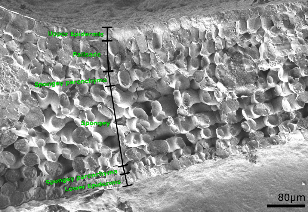
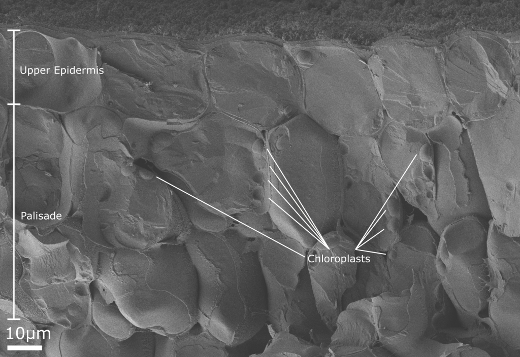
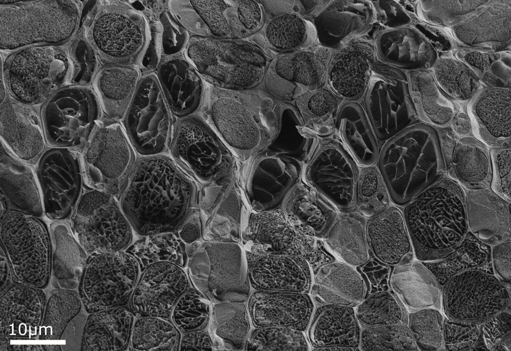
A freeze-fractured cryo-SEM image of a common privet leaf (Ligustrum vulgare) epidermis is shown above. It is possible to discern the distinct sections that make up the leaf structure. The microscope has resolution down to ~1nm, enabling it to capture the individual chloroplasts that produce the leaf’s energy through photosynthesis (image below). At the top of this image, the surface ornamentation of the leaf can also be seen.
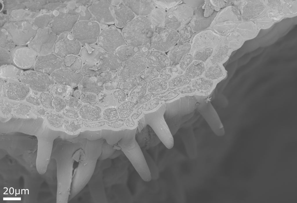
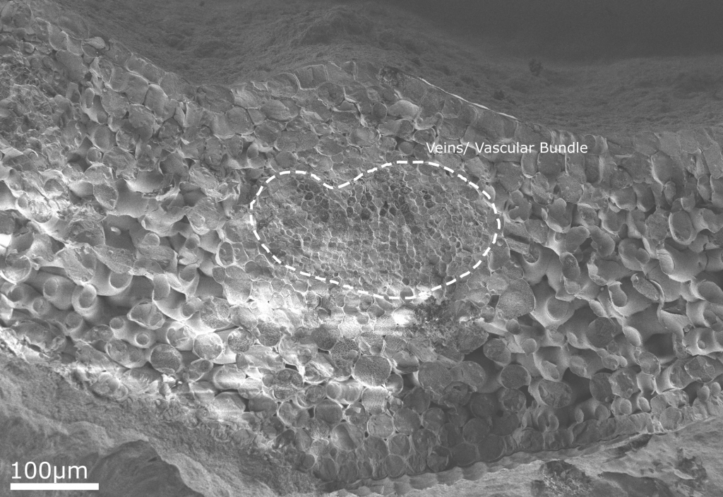
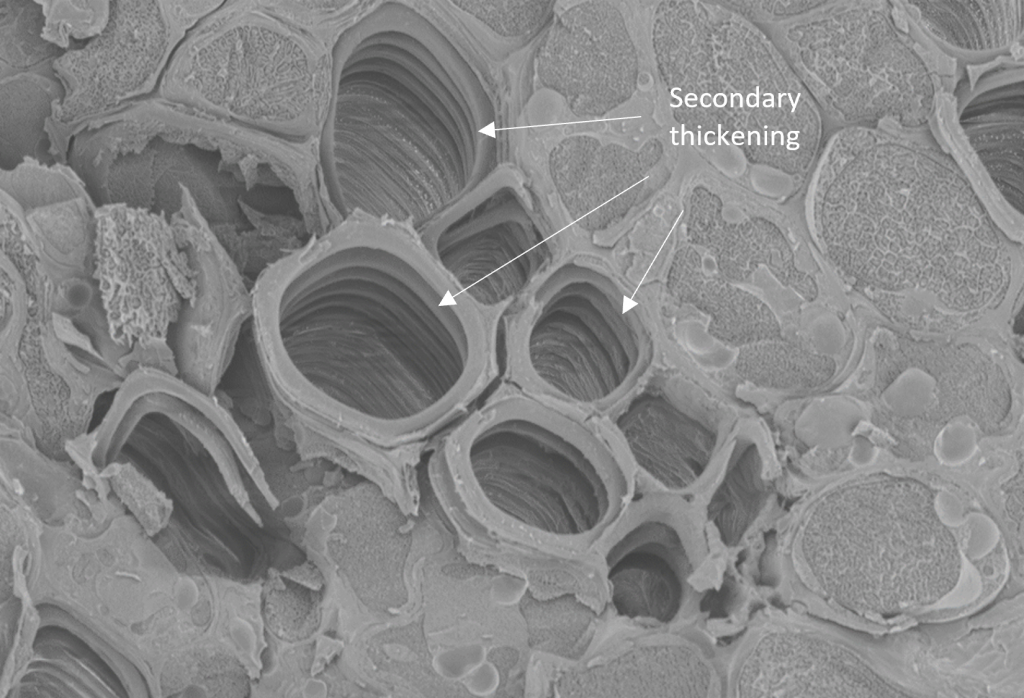
Another cross section of the leaf contains 1-2 layers of collenchyma cells (ones with thick primary walls). Below the epidermis, some hairs can be seen.
Please get in contact with us to find out more about ECFP and whether we can help you.
ECFP delivers fundamental product insight enabling improved formulation and processing for a more sustainable future.